


Absence of the Septum Pellucidum
Axial MRI, T1 sequence.

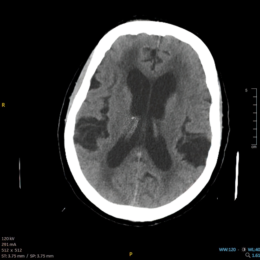
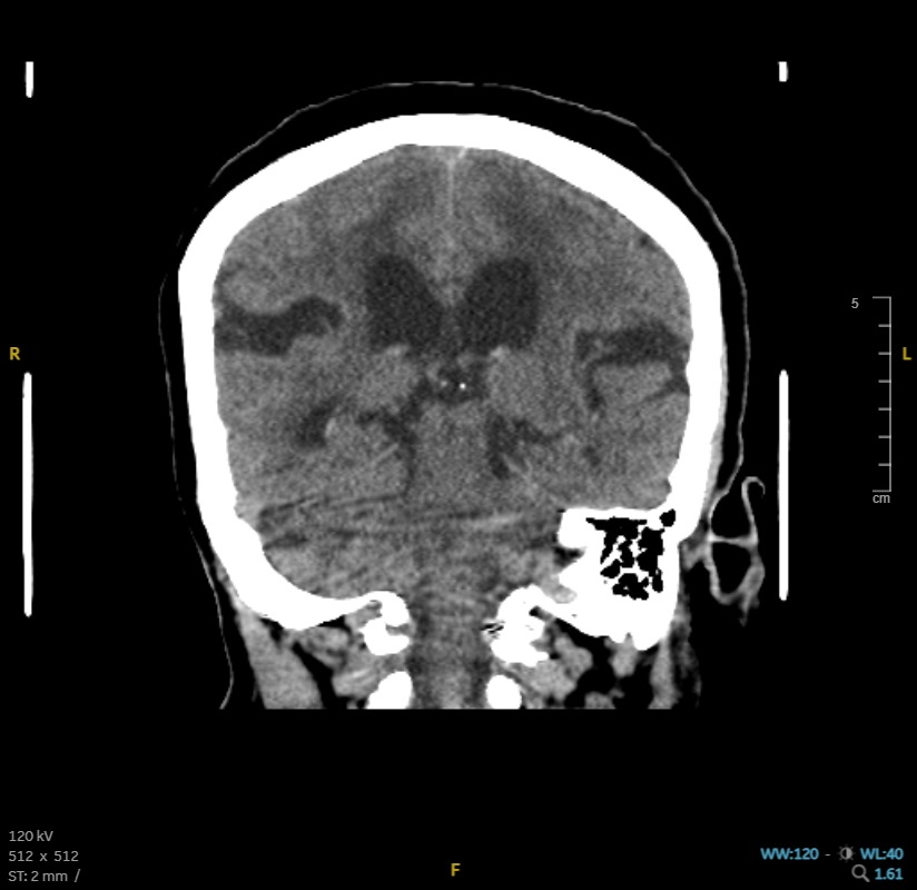
Aqueductal Stenosis
Axial head CT showing severe obstructive hydrocephalus due to aqueductal stenosis.

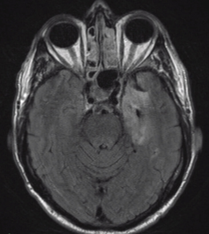
Central Pontine Myelinolysis
T2 FLAIR, axial MRI brain. Note the hyperintensity with the pons.
")
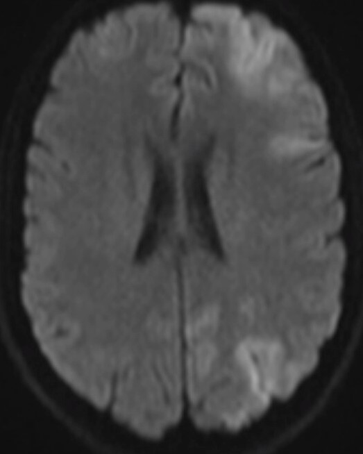
Creutzfeldt-Jakob Disease (CJD)
MRI DWI, axial cut, showing cortical ribboning.
")
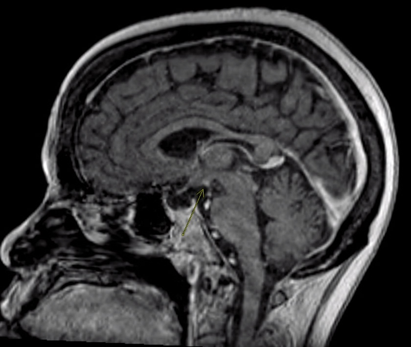
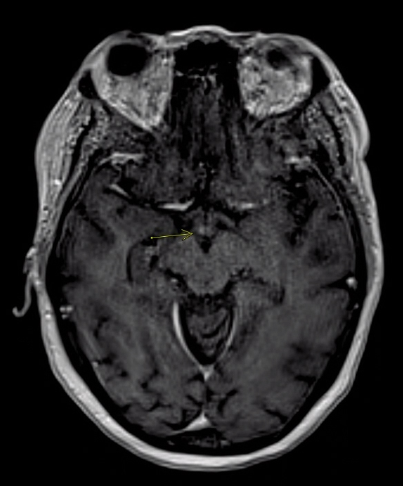
Wernicke's Encephalopathy (WE)
Axial MRI, T2 FLAIR sequence. Hyperintensity within the bilateral midbrain tectum is seen, which is classic of WE.

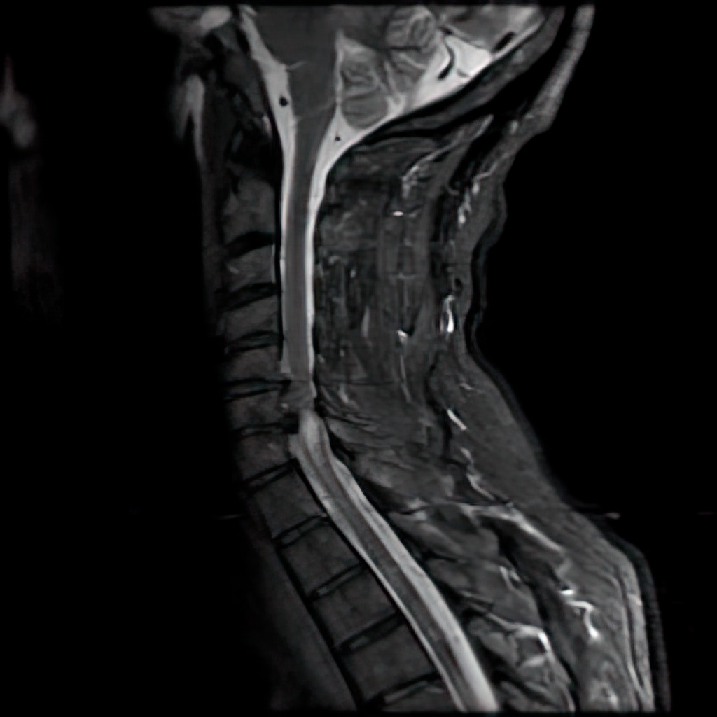
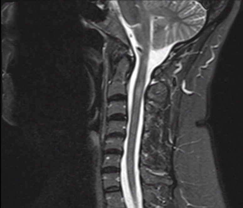
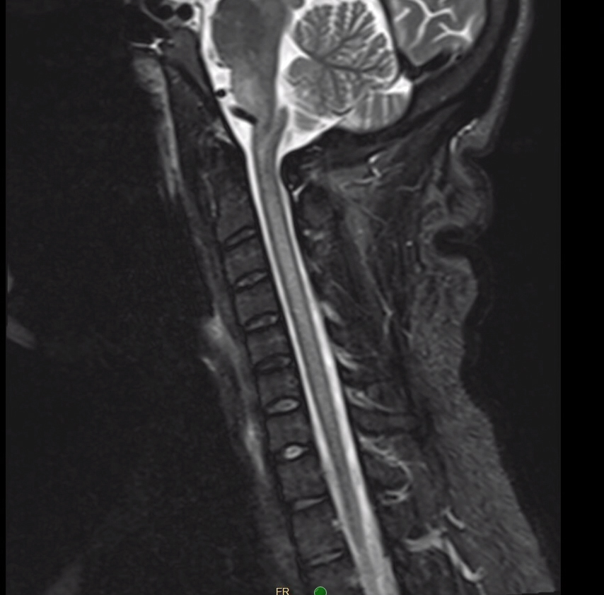
Spinal Syrinx and Chiari Malformation
Sagittal T2-weighted cervical spine MRI. Note the cavity within the cord that is isointense relative to the CSF. Syringomyelia (syrinx) is sometimes seen alongside Type 1 Chiari malformation.

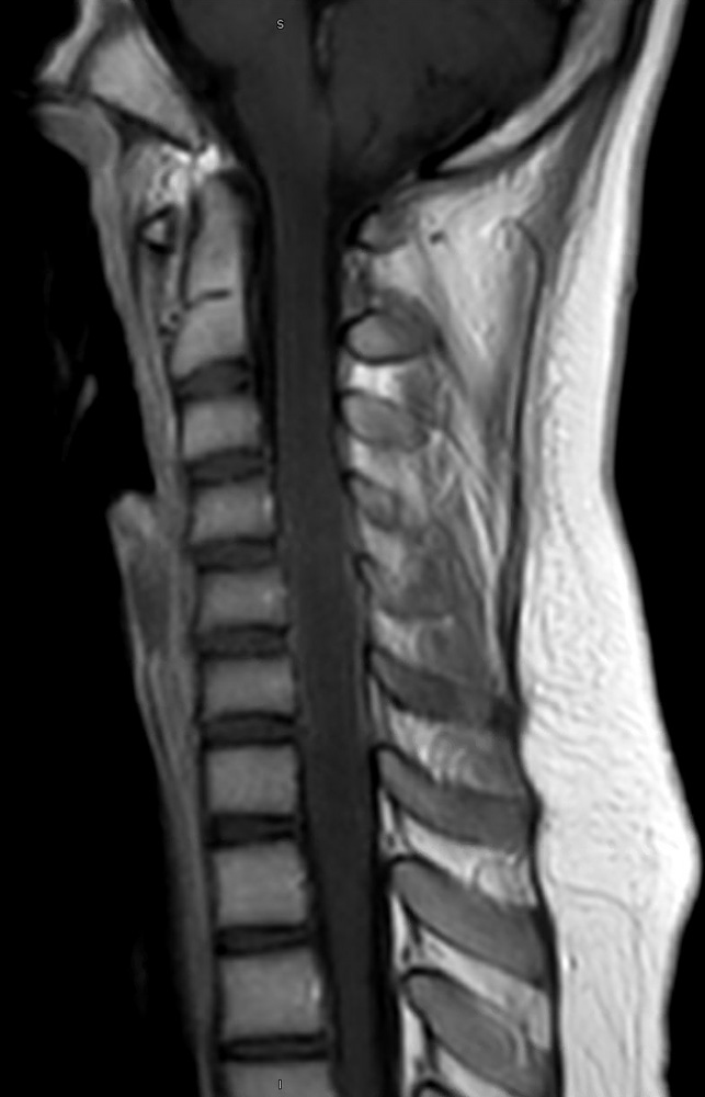
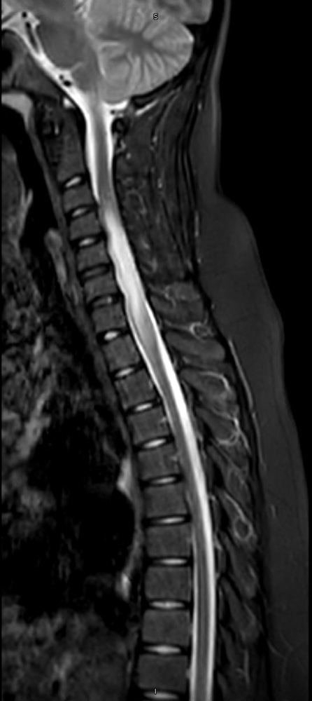
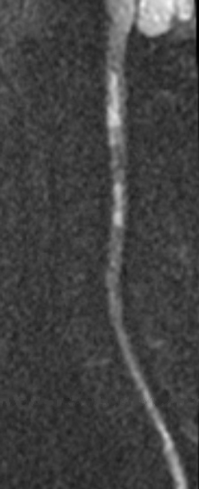
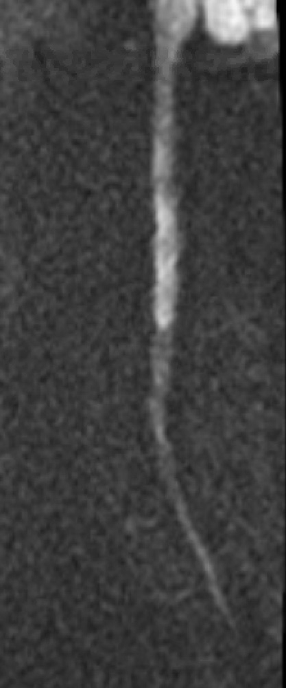
Multiple Sclerosis
Left: Sagittal spine MRI, T1 sequence. Middle: T1 w/ contrast. Right: T2. Note the contrast-enhancing cervical lesion, consistent with an active plaque.

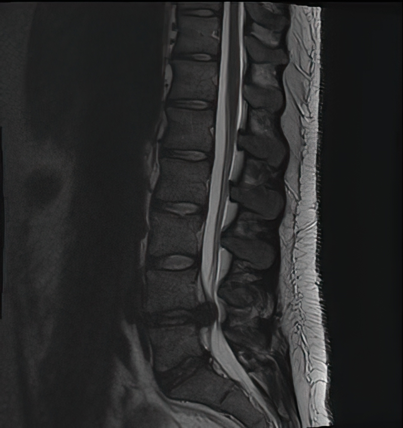
Guillain-Barre Syndrome
Axial spine MRI with contrast. Note the contrast enhancement of the cauda equina and nerve roots.

Odontoid Fracture
Cervical spine, coronal view.

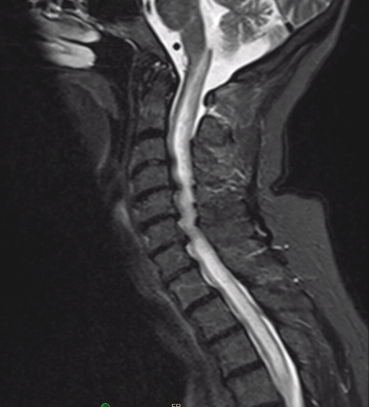
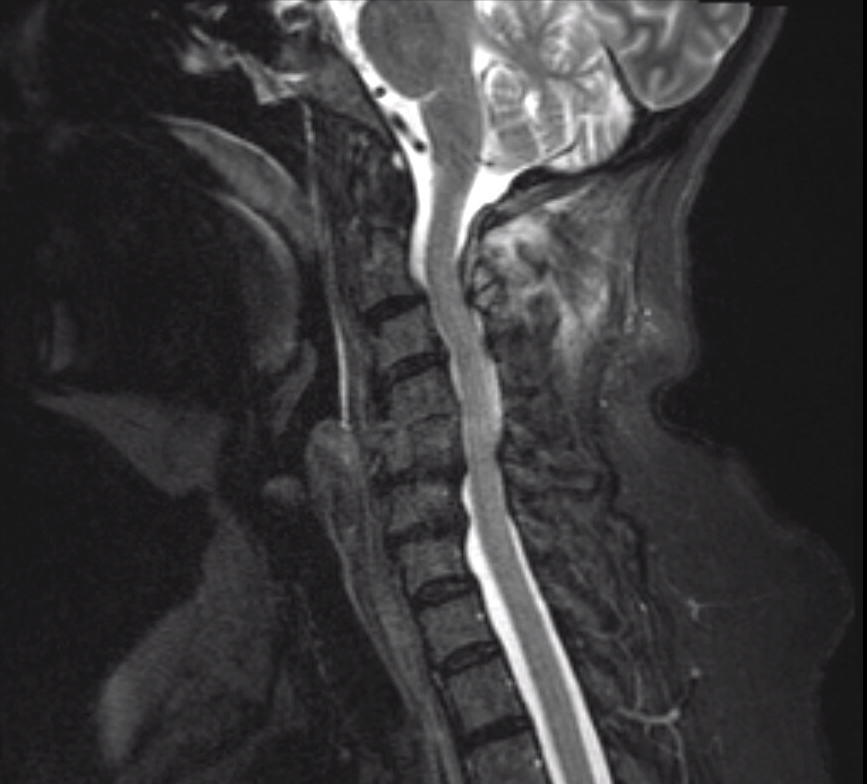
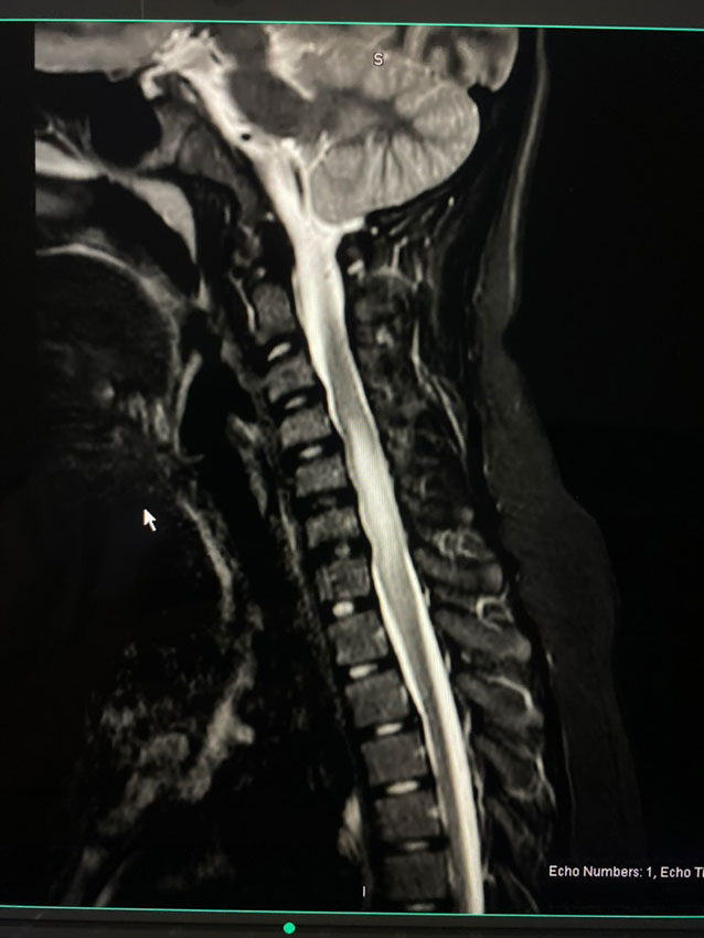
Spinal Cord Contusion
MRI cervical spine, sagittal view. The contusion is secondary to cervical spine fracture.

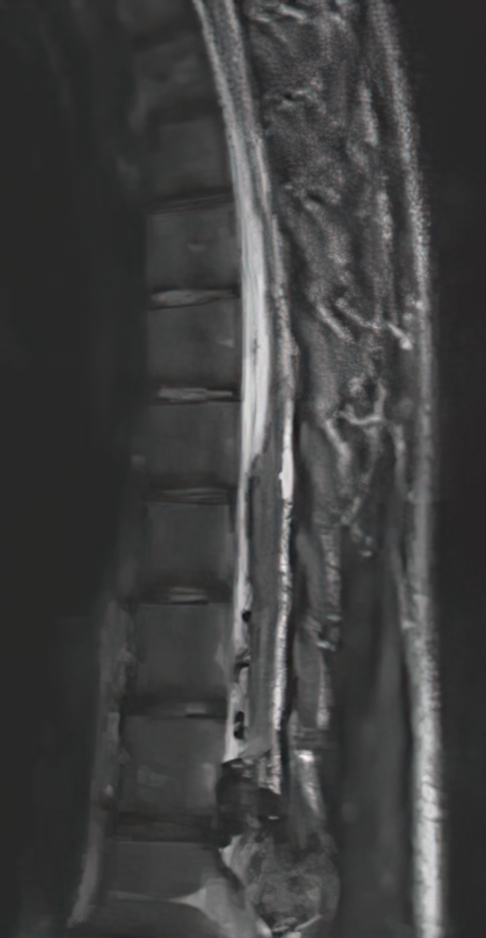
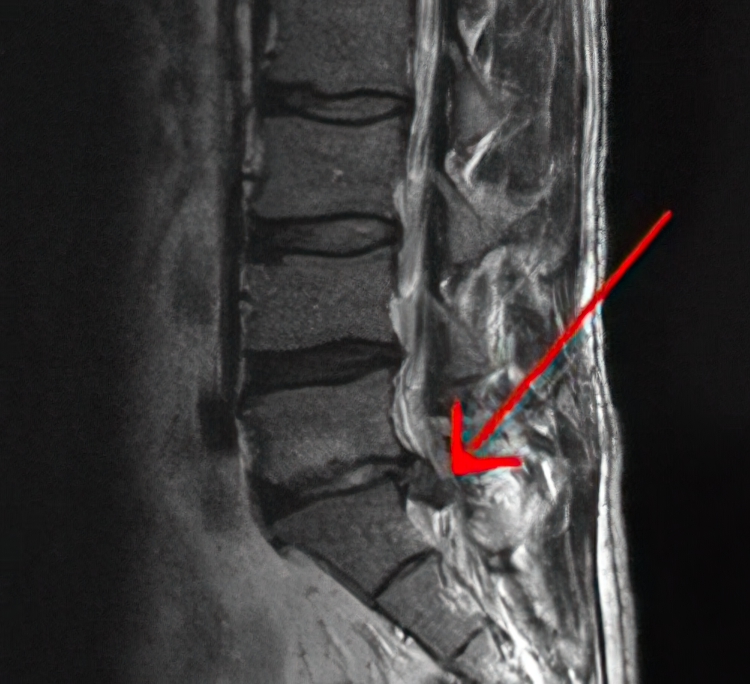
Lumbar spine MRI showing a destructive sacrococcygeal lesion suggestive of chordoma
Lumbar spine MRI, sagittal view.

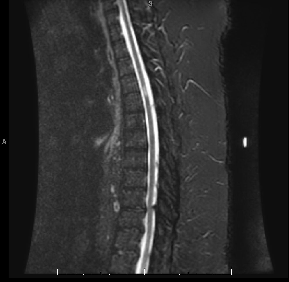
Metastatic Tumor to the Spine
Lumbar spine MRI, sagittal view.

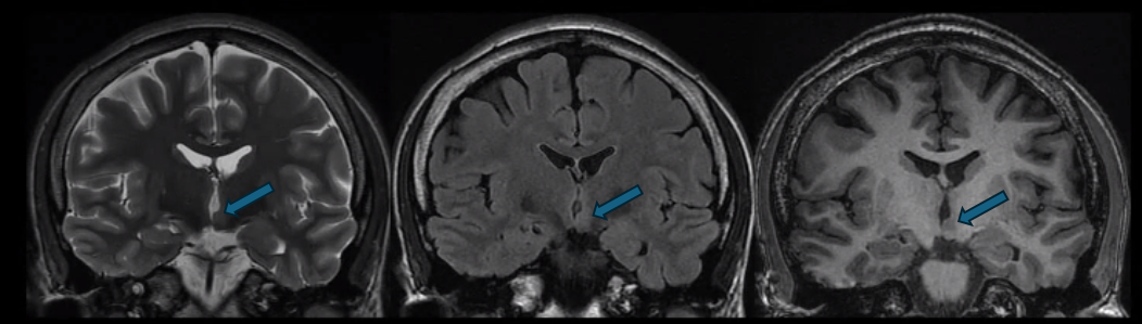
Left Mesial Temporal Sclerosis
Coronal MRI, T2 sequence.

Type 1 Odontoid Fracture
Left: Axial CT head. Right: Coronal.

Adrenoleukodystrophy
Axial T2 FLAIR MRI showing posterior-predominant white matter hyperintensities consistent with adrenoleukodystrophy.

Schizencephaly
Axial MRI, T2 FLAIR, showing open-lipped (Type 2) schizencephaly with unfused edges and exposure to the subarachnoid space.

Agenesis of the Corpus Callosum
Sagittal T2-weighted MRI showing agenesis of the corpus callosum.

Ventriculomegaly
Sagittal T2 FLAIR MRI showing severe ventriculomegaly in a 37-week-old patient.

Focal Cortical Dysplasia
Axial T2 FLAIR MRI showing focal hyperintensity of the right parietal cortex.

Nodular Heterotopia
Coronal T1 MRI showing periventricular grey matter.

Nodular Heterotopia
Coronal MRI, T2 sequence, showing periventricular grey matter (heterotopia).
")
Right Arteriovenous Malformation (AVM)
T2 brain MRI, axial cut.

Basilar Occlusion
Axial CTA.

Right Epidural Hematoma
Axial CT head.

Intracerebral Hemorrhage of Left Thalamus
This was secondary to hypertension. Note the intraventricular spread.

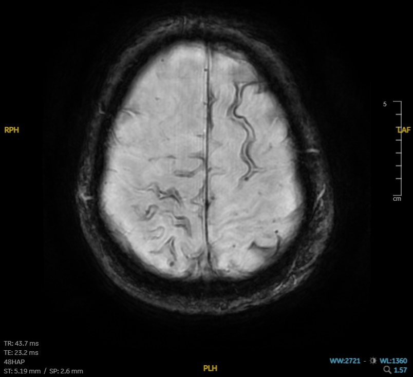
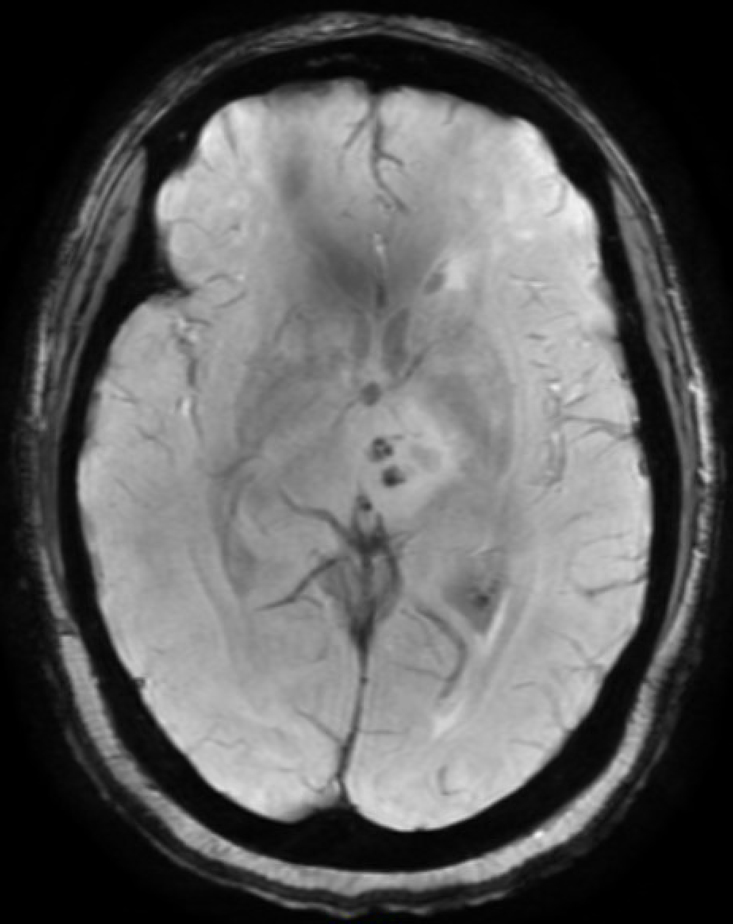
Cerebral Amyloid Angiopathy
Axial MRI GRE with numerous micro-hemorrhages.

Left Hyperdense MCA Sign
Axial CT.
.")
Hereditary and Metabolic Disorders Discussion: This clinical picture describes a typical presentation and MRI findings of Joubert syndrome; in which there is hypoplasia of the cerebellar vermis. The most common features of this syndrome include hyperpnea, hypotonia, oculomotor apraxia, ataxia, and intellectual disability. Other neurologic manifestations include seizures. The molar tooth sign, which is the result of the thickening and horizontalization of the superior cerebellar peduncle and a deep interpeduncular fossa, can also be seen in several other disorders including Dekaban-Arima syndrome, Senior-Loken syndrome, and COACH (cerebellar vermis hypoplasia, oligophrenia, ataxia, coloboma, and hepatic fibrosis).
Hereditary and Metabolic Disorders
Discussion:
This clinical picture describes a typical presentation and MRI findings of Joubert syndrome; in which there is hypoplasia of the cerebellar vermis. The most common features of this syndrome include hyperpnea, hypotonia, oculomotor apraxia, ataxia, and intellectual disability. Other neurologic manifestations include seizures. The molar tooth sign, which is the result of the thickening and horizontalization of the superior cerebellar peduncle and a deep interpeduncular fossa, can also be seen in several other disorders including Dekaban-Arima syndrome, Senior-Loken syndrome, and COACH (cerebellar vermis hypoplasia, oligophrenia, ataxia, coloboma, and hepatic fibrosis).

Multiple Meningiomas in NF Type 2
Axial MRI, T1 w/ contrast.

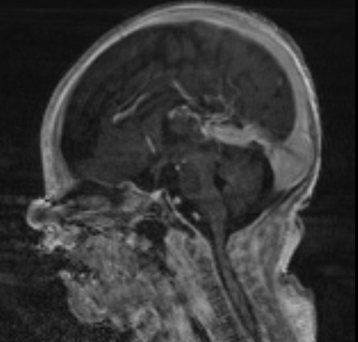
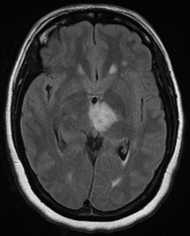
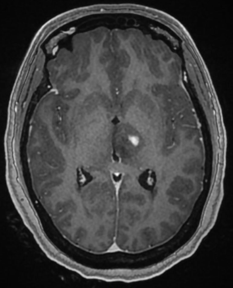
Germinoma
Left: Sagittal MRI, T2 FLAIR. Right: T1 w/ contrast. Note the mass in the pineal region.

Brain Metastasis
Axial CT without contrast, showing lesion with edema.

Dawson's Fingers in Multiple Sclerosis
Sagittal T2 FLAIR MRI.
