

Embryology and developmental anatomy can be complex topics, but it is important to understand the basics to translate to disease pathology and to do well on neurology exams. This chapter review contains helpful text and diagrams, and a practice quiz.
Authors: Matthew Ginsberg MD, Brian Hanrahan MD
Chapter Multimedia Content
Table of Contents
Table of Contents
Neural development
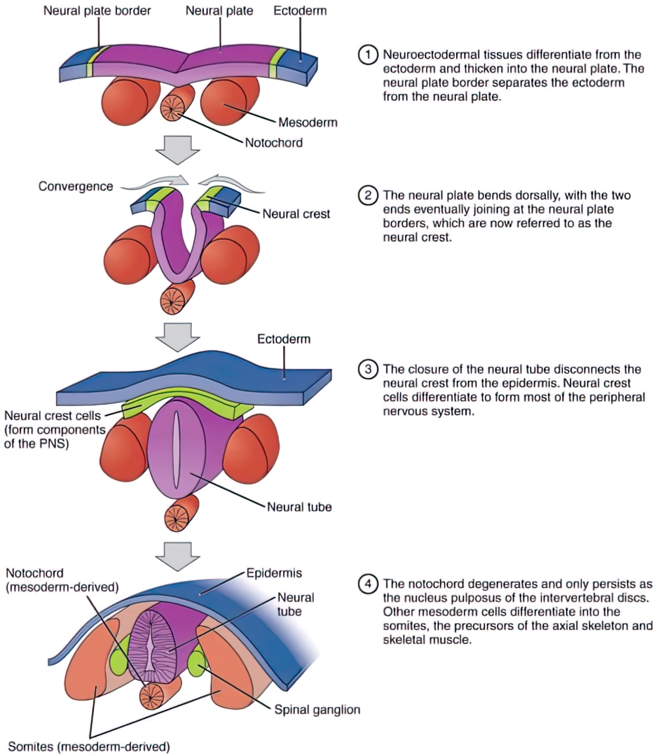
- All neuronal tissue is derived from the ectoderm germ layer.
- The notochord (mesoderm) induces the overlying neuroectoderm to differentiate into the neural plate at 18 days gestation.
- The notochord becomes part of the intervertebral discs in final development
- During the 3rd-5th weeks of gestation, the neural plate invaginates to form the neural tube (Figure 1).
- Neurulation occurs in two phases: Primary neurulation (dorsal induction) and secondary neurulation (caudal induction).
- Defects of primary or secondary neurulation will lead to various cortical and spinal cord malformations.
- The neural tube develops into the brain, brainstem, spinal cord, and preganglionic sympathetic neurons.
- During this time the fetus is most at risk for neural tube deficits (NTD).
- Folate deficiency, valproic acid, and carbamazepine are known to cause NTDs, especially spina bifida.
- The ventral aspect of the neural tube is the basal plate.
- The dorsal aspect of the neural tube is the alar plate.
- Neurulation occurs in two phases: Primary neurulation (dorsal induction) and secondary neurulation (caudal induction).
- By week 10, the basal plate (ventral) of the neural tube becomes motor neurons of the cranial nerve and anterior horn gray matter while the alar plate (dorsal) becomes posterior horn gray matter and sensory neurons.
- Neural crest cells that border the neural tube lead to the growth of the peripheral nervous system: Schwann cells, neuroglial cells, parasympathetic, and postganglionic sympathetic fibers.
- Neural crest cells also develop into melanocytes, pia and arachnoid, thyroid, and teeth.
Figure 1: Early Embryonic Development of Nervous System
Breakdown of the developing brain
- There are three primary vesicles of the developing brain at 4 weeks:
- Forebrain (prosencephalon): Develops into the telencephalon and diencephalon.
- Midbrain (mesencephalon)
- Hindbrain (rhombencephalon): Develops into the metencephalon and myelencephalon.
- By 5 weeks, these develop into five secondary vesicles:
- Telencephalon: Develops into the cerebral hemispheres, basal ganglia, hippocampus, and amygdala.
- Diencephalon: Develops into the thalamus, hypothalamus, and retina.
- Mesencephalon: Develops into the midbrain.
- Metencephalon: Develops into the pons and cerebellum.
- Myelencephalon: Develops into the medulla.