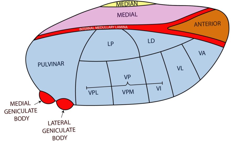
The thalamus and hypothalamus are complicated subcortical structures. While they both originate from the diencephalon, these two structures have starkly different roles within the central nervous system. The thalamus is a subcortical grey matter structure that acts as a major relay center between the cortex and other subcortical areas while the hypothalamus has many nuclei with various roles; hormone synthesis, temperature regulation, hunger/thirst, etc. The hypothalamic-pituitary axis is an important topic for introductory neuroanatomy courses but is considered low yield for in-service and board examinations. Because of this, the intricacies of this topic will not be covered in-depth in this chapter. The goal of this chapter is to review the complicated roles of the thalamus and associated pathology and briefly review hypothalamic nuclei.
Author: James Eaton, MD
Editor: Brian Hanrahan, MD, Steven Gangloff MD
Thalamus
- The thalamus is a collection of subcortical grey matter and part of the diencephalon (along with the hypothalamus) involved in relaying information between different functional regions of the CNS.
- It borders the dorsal part of the third ventricle and functions as part of the lateral wall of the ventricle.
Thalamic nuclei
- There are three main types of thalamic nuclei: relay, association, and nonspecific nuclei.
- Relay nuclei have very distinct inputs and outputs.
- Association nuclei receive a majority of their input from the cortex and project fibers to other cortical regions.
- Nonspecific nuclei broadly project to numerous cortical regions.
Relay nuclei
- Ventral posterior lateral (VPL) nucleus: Relay center for sensory information of the body. The output is to the postcentral (sensory) gyrus.
- Ventral posterior medial (VPM) nucleus: Relay center for sensory information of the face. The output is to the postcentral (sensory) gyrus.
- The VPM is also the relay center for taste.
- Lateral geniculate body/nucleus (LGN): Relay center for vision. Receives input from the retina with output to the primary visual cortex via optic radiations.
- Optic radiations from the upper visual world travel through the temporal lobe and optic radiations from the lower visual world pass through the parietal lobe.
- Medial geniculate body/nucleus (MGN): Relay center for hearing. Receives input from the inferior colliculi and projects to the primary auditory cortex.
- Ventral anterior (VA) nucleus: Conveys motor information from basal ganglia to the premotor cortex. It plays a role in the initiation of movement.
- Ventral lateral (VL) nucleus: Conveys motor information from both the cerebellum and basal ganglia to the primary motor cortex.
Log in to View the Remaining 60-90% of Page Content!
New here? Get started!
(Or, click here to learn about our institution/group pricing)1 Month Plan
Full Access Subscription-
Access to all chapters
-
Access to all images and cases
-
Access to all flashcards
-
Access to Full Question Bank
3 Month Plan
Full Access Subscription-
Access to all chapters
-
Access to all images and cases
-
Access to all flashcards
-
Access to Full Question Bank
1 Year Plan
Full Access Subscription-
Access to all chapters
-
Access to all images and cases
-
Access to all flashcards
-
Access to Full Question Bank


