

- The majority of the cortex has 6 cortical layers
- The six layers from superficial to deep:
- Molecular
- External granular
- External pyramidal
- Internal granular
- Internal pyramidal (ganglion)
- Multiform.
- The six layers from superficial to deep:
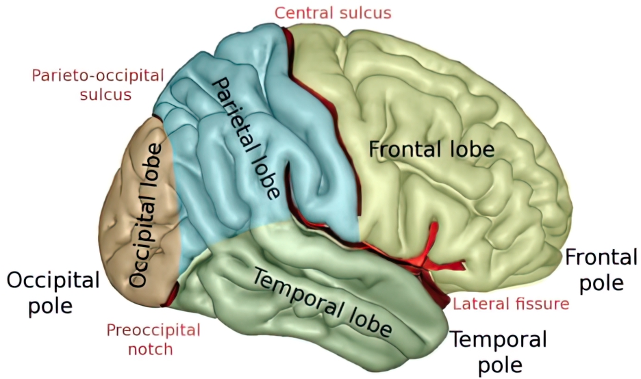
- The cortex is organized into 5 lobes: frontal, parietal, temporal, occipital, and limbic.
- Frontal lobe: Bound by the central sulcus and lateral fissure.
- Parietal lobe: Bound by the central sulcus, lateral fissure, and parieto-occipital fissure.
- Temporal lobe: Bound by the Sylvian fissure and preoccipital notch.
- Occipital lobe: Bound by the parieto-occipital sulcus and preoccipital notch.
- Limbic lobe: Consists of the parahippocampal, cingulate, and subcallosal gyri. The limbic lobe interacts with various other structures that are part of the limbic system.

Lobes of the brain
- The majority of the cortex has 6 cortical layers
Frontal Lobe
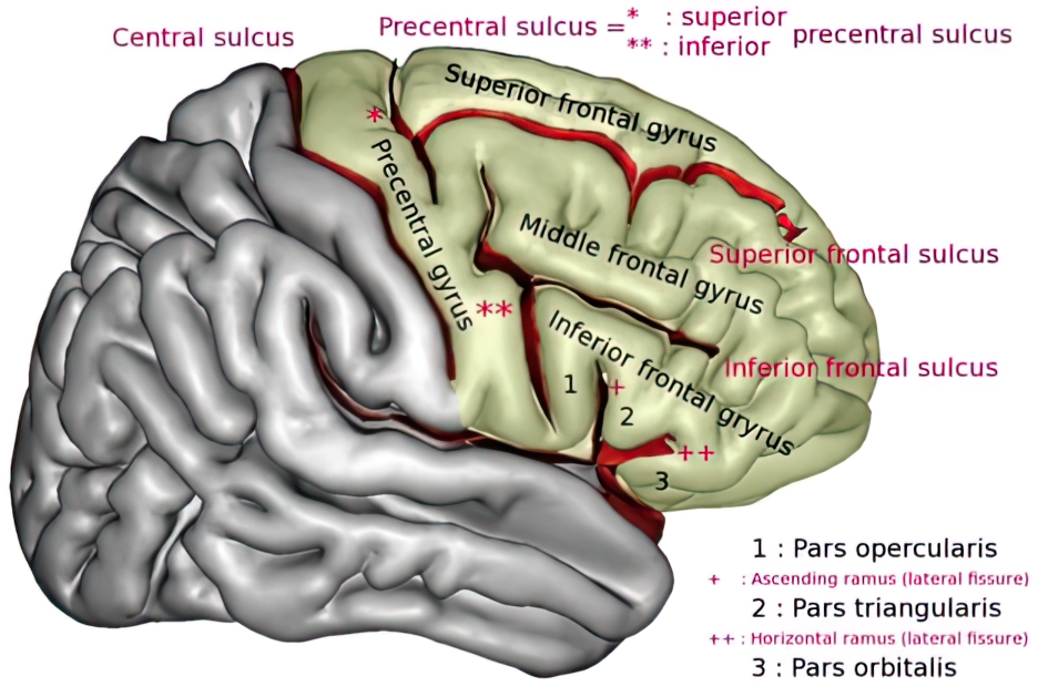
- There are four major gyri: precentral, superior frontal, middle frontal, and inferior frontal.
- The precentral gyrus is the primary motor strip.
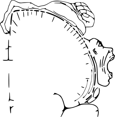
- The motor homunculus of the precentral gyrus:
- The superior frontal contains the supplementary motor area (SMA).
- The middle frontal contains the frontal eye fields which are necessary for voluntary saccadic eye movements.
- The inferior frontal is organized into the pars orbitalis, pars triangularis, and pars opercularis.
- Pars opercularis and triangularis are associated with Broca’s area.
- Pars orbitalis has a role in thought, cognition, and planning behavior.
Parietal Lobe
- There are five principal parts: post-central gyrus, superior parietal lobule, inferior parietal lobule, the precuneus, and the posterior portion of the paracentral lobule.
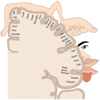
- The postcentral gyrus contains the primary sensory cortex.
- The sensory homunculus of the postcentral gyrus:
- The superior parietal lobule contains the somatosensory association area.
- The inferior parietal lobule has two components: angular and supramarginal gyri
- This is the sensory association cortex and has a role in perception, vision, reading, and speech.
- A lesion to this region can lead to Gerstmann’s syndrome.
- The precuneus is an area of cortex just anterior to the occipital lobe on the medial surface. It has a broad spectrum of functions including visuospatial processing, memory, and first-person perspective.
- It is an early region of atrophy in Alzheimer’s dementia
- The posterior portion of the paracentral lobule is regarded as a tertiary somatosensory cortex involved in stereognosis.
- Stereognosis: the perception, understanding, recognition, and identification of an object by touch.
- Tested by having the patient feel an object and identify it, such as a paperclip or set of keys.
- It is often accompanied by other deficits like agraphesthesia.
- Stereognosis: the perception, understanding, recognition, and identification of an object by touch.