Spine anatomy and pathology can be difficult, but it is an important part of neurology examinations including the boards and the RITE® exam. Here you will review vascular, traumatic, inflammatory, and other lesions of the cord and vertebrae using high-yield images, text, and practice questions.
The vertebral arteries and the aorta (via 10 medullary branches) provide the primary vascularization of the spinal cord by forming a single anterior spinal artery (ASA) and two posterior spinal arteries (PSA).
The largest medullary branch, the great anterior artery of Adamkiewicz, arises between T9 and L2 and supplies the lumbar enlargement.
The ASA supplies the anterior two-thirds of the spinal cord, and the two PSAs together supply the posterior one-third.
The upper thoracic (T1-T4) segments are in the zone between ascending and descending blood supply are thus act as a watershed vulnerable to ischemic insult by hypoperfusion/hypotension.
Nerve root anatomy
Cervical nerve roots exit above the corresponding vertebrae and the C8 root exists above the T1 vertebrae.
Thoracic, lumbar, and sacral nerve roots exit below their corresponding vertebral body, laterally and superiorly through the neural foramina.
Therefore, a herniation at the L4-L5 disc interspace will compress the L4 nerve root as it exits.
Autonomic dysfunction can be an additional complication of spinal lesions above T6, and this is one of the leading causes of mortality in this population.
It presents with respiratory distress, impaired thermoregulation, lower urinary tract and GI complications, and cardiovascular dysfunction such as autonomic dysreflexia.
Autonomic dysreflexia is sudden episodic increases in blood pressure along with baroreceptor-mediated bradycardia in response to noxious visceral or cutaneous stimulation below the injury level.
Motor and sensory pathways
Efferent (Motor) Pathway
The corticospinal tract sends motor instruction from the pre-central gyrus to muscles of the contralateral side after decussation in the pyramids of the medulla.
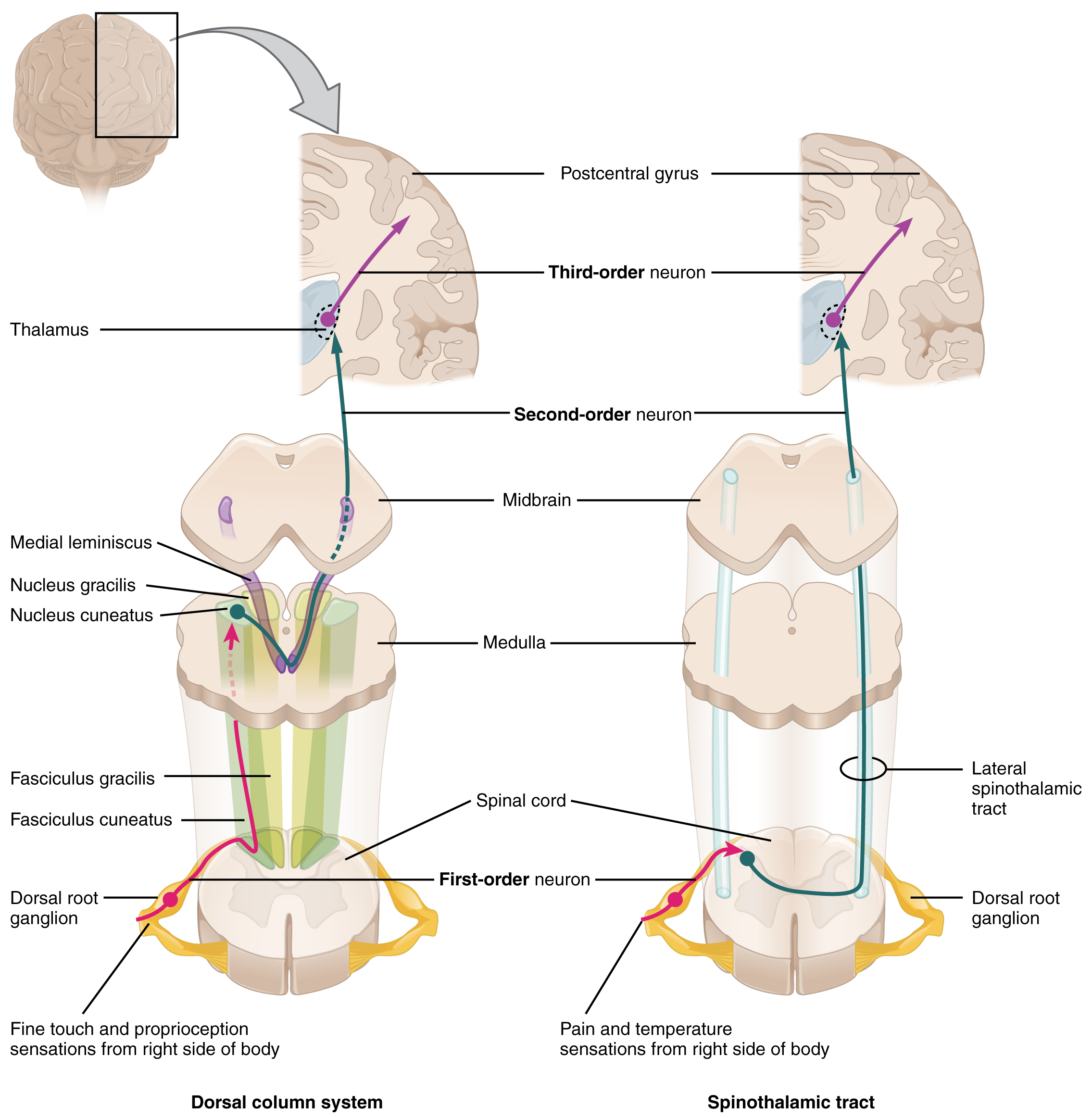
Afferent (Sensory) Pathways
Left: Dorsal column system receiving fine touch and proprioception input and delivering to the cortex after decussation in the medulla. Right: Spinothalamic tract receiving pain and temperature sensation and delivering to the cortex after decussation within the spinal cord.
Spinal Cord Anatomy and Lesions
A cursory understanding of spinal cord anatomy will allow you to interpret the presenting features of diseases that target certain structures.
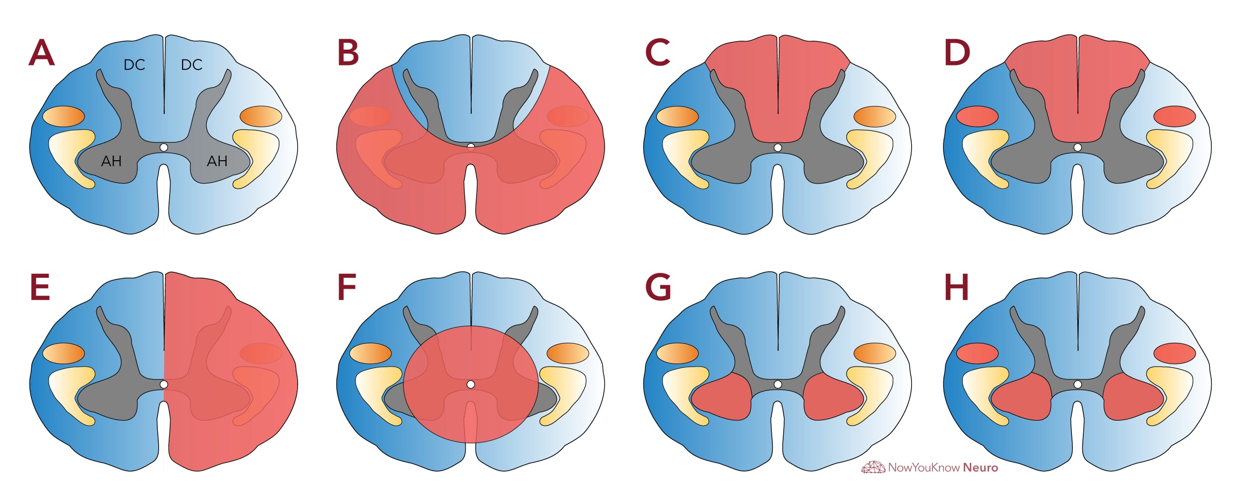
The diagram below includes commonly tested partial spine lesions and their anatomic correlates.
The anterior horn (AH) and corticospinal tracts (orange) relay efferent motor output. The dorsal columns (DC) transmit afferent fine touch and proprioception input to the brain. The spinothalamic tract (yellow) relays afferent pain and temperature sensation.
A: Normal spinal cord. Anterior horn (AH) and lateral corticospinal tracts (orange) relay efferent motor output. Dorsal columns (DC) transmit afferent fine touch and proprioception input to the brain. Spinothalamic tract (yellow) relays afferent pain and temperature sensation. B: Anterior cord syndrome/infarct. Damage marked in red. C: Posterior cord syndrome or tabes dorsalis of tertiary syphilis. D: Subacute combined degeneration from B12 deficiency. E: Brown-Sequard cord hemisection. F: Central cord syndrome or syrinx. G: Poliomyelitis. H: Amyotrophic lateral sclerosis.
Log in to View the Remaining 60-90% of Page Content!
New here? Get started!
(Or, click here to learn about our institution/group pricing)
Our “Board Pass Guarantee” is designed to provide added confidence and support for users preparing for the ABPN “Initial Certification in Neurology” or ABPN “Continuing Certification in Neurology” examinations. The following terms and conditions apply:
Eligibility
The Board Pass Guarantee is only available to users who purchase a 3-month or 1-year subscription to our platform.
To qualify for the guarantee, users must complete at least 50% of the question bank associated with their account before the date on which they took the ABPN exam.
To qualify for the guarantee, users must have taken the ABPN board exam within 14 months of the purchase of their NowYouKnowNeuro account.
This guarantee is effective only for Board exams taken after 07/01/2024.
Guarantee Benefits
If a user fails their ABPN “Initial Certification in Neurology” or ABPN “Continuing Certification in Neurology” examination, they will receive an account extension equal to the duration of their original subscription (either 3 months or 1 year).
Proof of Eligibility
To claim the guarantee, users must submit an image or screenshot of their official ABPN failure letter. The document must clearly indicate the user’s name, the exam taken, and the result.
Limitations
This guarantee does not provide refunds for prior purchases.
The guarantee does not cover any fees or costs associated with taking the ABPN exam, including but not limited to registration fees, travel expenses, or other study materials.
This guarantee does not apply to other exams beyond the ABPN “Initial Certification in Neurology” or ABPN “Continuing Certification in Neurology” examinations.
This guarantee may be used once per person.
How to Claim
To request an account extension under the Board Pass Guarantee, users must contact our support team via our Contact Us form within 30 days of receiving their official ABPN failure letter. We will ask for the required proof of eligibility via email as outlined above.
General Terms
The Board Pass Guarantee is subject to verification and approval by our team. Misrepresentation or submission of falsified documents will result in disqualification from the guarantee and may lead to account suspension.
By participating in the Board Pass Guarantee, users agree to these terms and conditions, which are subject to change at our discretion.