

In this chapter, we will be covering the vascular anatomy of the brain, brainstem, and spinal cord. Additionally, we will be covering typical stroke presentations. After reviewing this chapter, one will (1) better understand the anterior and posterior cerebral vascular systems, (2) be able to identify stroke syndromes based on the neurological deficit, and (3) be able to identify vascular territories on imaging.
If you want to learn more about the pathophysiology and management of stroke check out our Vascular Neurology chapter.
Author: Brian Hanrahan MD
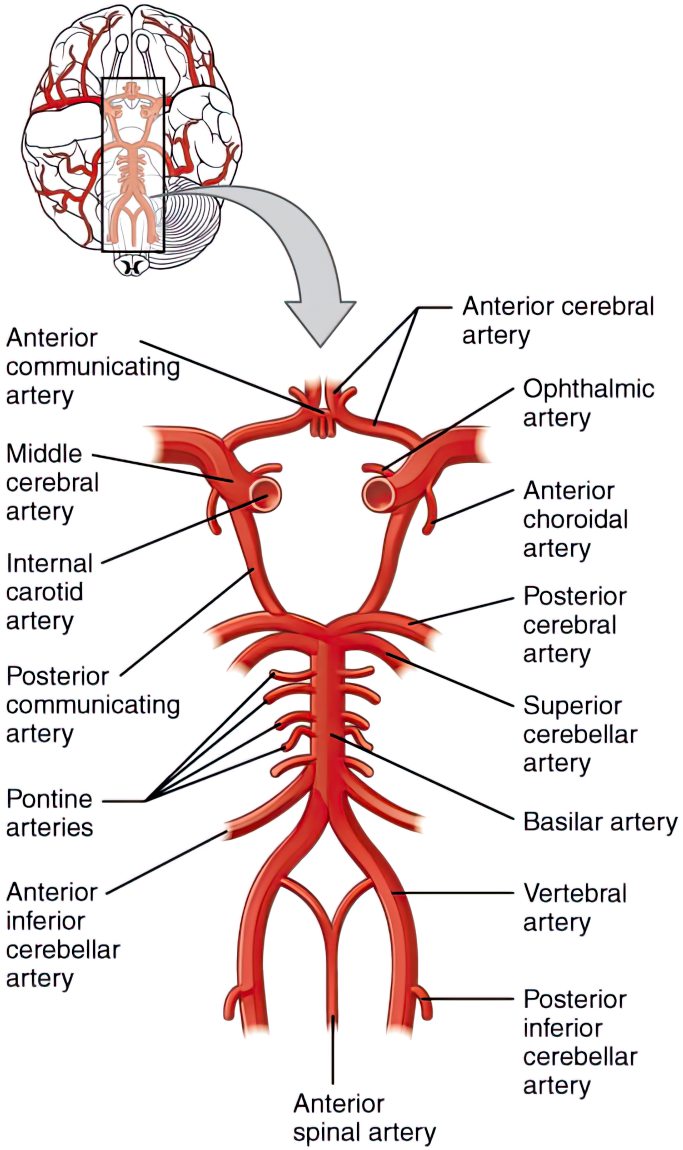
Figure 1: The Cerebral Vascular System
Introduction
- The cerebral vascular system can be subdivided into anterior and posterior circulatory systems.
- Two internal carotid arteries (ICAs) provide the anterior circulation.
- The vertebral arteries/basilar artery provides the posterior circulation.
- Cerebral angiogram (DSA) provides the best imaging quality to identify any vascular pathology.
- CT angiograms and MRA imaging can also be used to evaluate vascular anatomy but is less sensitive than a cerebral angiogram.
Note:
Anterior circulation strokes are much more common than posterior circulation strokes
Anterior Circulation
- The left common carotid artery branches off the aortic arch while the right common carotid artery branches off the brachiocephalic artery.
- The common carotid artery divides into external and internal carotid arteries at the level of the fourth cervical vertebra.
- The external carotid supplies structures in the neck, face, and meninges.
- The internal carotid supplies the anterior portion of the brain, eye, optic nerve, anterior pituitary, and meninges.
- The common carotid artery divides into external and internal carotid arteries at the level of the fourth cervical vertebra.
Reminder
The middle meningeal artery, which enters the skull cavity via the foramen spinosum, is a distal branch of the external carotid artery. NOT the internal carotid artery!
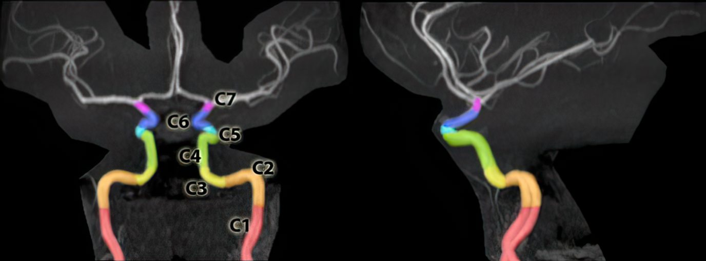
- The internal carotid artery has seven segments (according to the Bouthillier Classification):
Add terminal branches
- Cervical (C1)
- Petrous (C2)
- Lacerum (C3)
- Cavernous (C4)
- Clinoidal (C5)
- Ophthalmic (C6)
- Communicating (C7)
- Cervical (C1)
- Petrous (C2)
- Caroticotympanic artery
- Vidian artery
- Lacerum (C3)
- Cavernous (C4)
- Meningohypophyseal trunk
- Inferolateral trunk
- Capsular arteries of McConnell (variable)
- Clinoidal (C5)
- Ophthalmic (C6)
- Ophthalmic artery
- Superior hypophyseal artery
- Communicating (C7)
- Posterior communicating artery (PCOM)
- Anterior choroidal artery
- Anterior cerebral artery (ACA)
- Middle cerebral artery (MCA)
(Note: Gibo and Fisher Classifications number these slightly different)
Log in to View the Remaining 60-90% of Page Content!
New here? Get started!
(Or, click here to learn about our institution/group pricing)1 Month Plan
Full Access Subscription
$142.49
$
94
99
1 Month -
Access to full question bank
-
Access to all flashcards
-
Access to all chapters & site content
3 Month Plan
Full Access Subscription
$224.98
$
144
97
3 Months -
Access to full question bank
-
Access to all flashcards
-
Access to all chapters & site content
1 Year Plan
Full Access Subscription
$538.47
$
338
98
1 Year -
Access to full question bank
-
Access to all flashcards
-
Access to all chapters & site content
Popular
Table of Contents
Loading table of contents...