
Tau protein
- Function: Assists in microtubule function as a microtubular associated protein.
- Genetics: encoded by microtubule-associated protein tau (MAPT) gene
- Pathology: Cell death leads to leakage of cellular contents leading to elevated levels of tau and formation of neurofibrillary tangles which are immunoreactive for phosphorylated tau.
Alzheimer’s disease (AD)
- Presents with impairment of memory and at least one other area of cognition.
- Early symptoms include forgetfulness for recent events or newly acquired information, disorientation, and difficulty with complex cognitive functions.
- The degree of memory loss usually correlates with the severity of the loss of cholinergic neurons in the nucleus basalis of Meynert, which has cholinergic projections to the cerebral cortex.
Genetics
- Amyloid precursor protein (APP):
- Located on chromosome 21.
- People with Down syndrome (trisomy 21) have a higher risk of Alzheimer’s disease due to the overproduction of APP.
- Amyloid precursor protein (APP):
- Apolipoprotein E (APOE) gene:
- Susceptibility gene found on chromosome 19.
- Associated with late-onset Alzheimer disease (AD)
- Works by assisting the metabolism and transport of β-amyloid.
- APOE 4 allele increases risk by 5-15 fold.
- APOE 2 allele decreases the risk of AD.
Biomarkers
- Used to help predict conversion from MCI to AD.
- CSF studies: Can be more sensitive than cognitive testing to predict conversion to AD from MCI.
- Elevated total and phosphorylated tau levels due to dying and damaged neurons
- Decreased beta-amyloid 1-42 due to an accumulation of amyloid in plaques.
Pathology
- Microscopic analysis will show extracellular amyloid neuritic plaques and dystrophic neurites with reactive astrocytes and microglia.

Neuritic Plaques
Extracellular amyloid plaque on silver stain, high power.

Alzheimer’s Disease
Amyloid stain with beta-amyloid plaque (low magnification).

Neuritic Plaques
On silver stain.
- Intracellular neurofibrillary tangles, Hirano bodies, neuronal granulovacuolar degeneration, and the deposition of amyloid in the walls of blood vessels can also be seen.
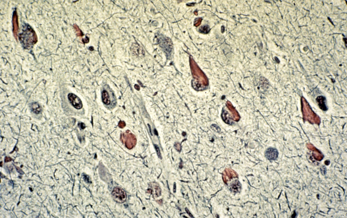
- Neurofibrillary tangles: Flame-shaped intracellular inclusion bodies of phosphorylated tau protein.
- Hirano bodies: Intracellular aggregates of actin and actin-associated proteins, which are not specific to Alzheimer’s disease.
- Granulovacuolar degeneration: Intraneuronal accumulation of large (up to 5 µm diameter) double membrane-bound vacuoles harboring a central granule.

Alzheimer’s Disease
Neurofibrillary tangles on H&E stain.

Granulovacuolar degeneration
High power H&E microscope image showing intraneuronal large vacuoles (arrows), each harboring a central granule. Seen with granulovacuolar degeneration of Alzheimer’s disease.

Granulovacuolar degeneration
Medium power H&E image showing intraneuronal accumulations of large vacuoles harboring a central granule.

Alzheimer’s Disease
Tangles and plaques on lectin stain.

Alzheimer’s Disease
Neurofibrillary tangles on silver stain.

Alzheimer’s Disease
Stain showing a Hirano body.
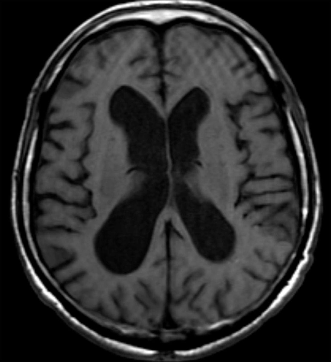
Imaging
- CT/MRI
- Widening of the cortical sulci and enlargement of the lateral ventricles
- Microhemorrhages secondary to amyloid angiopathy can also occur in cortical and subcortical locations.
- Hippocampal and high parietal atrophy